missing translation for 'onlineSavingsMsg'
Learn More
Learn More
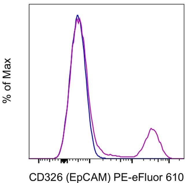
Invitrogen™ CD326 (EpCAM) Monoclonal Antibody (G8.8), PE-eFluor™ 610, eBioscience™, Invitrogen™

Rat Monoclonal Antibody
163.00 € - 376.00 €
Specifikationer
| Antigen | CD326 (EpCAM) |
|---|---|
| Klon | G8.8 |
| Konzentration | 0.2 mg/mL |
| Anwendungen | Flow Cytometry |
| Klassifikation | Monoclonal |
| Product Code | Brand | Menge | Price | Quantity & Availability | |||||
|---|---|---|---|---|---|---|---|---|---|
| Product Code | Brand | Menge | Price | Quantity & Availability | |||||
|
17104103
|
Invitrogen™
61-5791-80 |
25 μg |
163.00 €
25 Mikrogramm |
Log in om dit product te kopen Registreer vandaag om een webaccount aan te maken | |||||
|
17114103
|
Invitrogen™
61-5791-82 |
100 μg |
376.00 €
100 Mikrogramm |
Log in om dit product te kopen Registreer vandaag om een webaccount aan te maken | |||||
Description
The G8.8 MAb reacts with the 40 kDa protein mouse EpCAM (epithelial cellular adhesion molecule), also known as EGP40 (epithelial glycoprotein 40), 17-1A antigen, TACSTD1 (tumor-associated calcium signal transducer 1), and CD326. The immunogen used to generate the G8.8 antibody was the TE-71 thymic epithelial cell line. CD326 is expressed on the majority of epithelial cells, and is considered a pan-carcinoma antigen. CD326 mediates calcium-independent, homophilic, cell-cell adhesion and may function as a growth factor receptor. The antigen is being used as a target for immunotherapy treatment of human carcinomas. CD326 binds LAIR-1 (CD305) and LAIR-2 (CD306) to inhibit cellular activation and inflammation. This epithelial glycoprotein is now recognized as having an important role in tumor biology.
Ep-CAM (epithelial adhesion molecule, epithelial specific antigen, ESA) is a transmembrane glycoprotein expressed in the epithelium with a molecular weight of approximately 40 kDa, which functions as an epithelial cell adhesion molecule. Ep-CAM functions as a homotypic calcium-independent cell adhesion molecule, and has a direct impact on cell cycle, proliferation and metabolism of epithelial cells and fibroblasts due to its ability to rapidly induce the proto-oncogene c-myc and the cell cycle regulating genes cyclin A and E. Ep-CAM mediates Ca2+-independent homotypic interactions. Formation of Ep-CAM-mediated adhesions have a negative regulatory effect on adhesions mediated by classic cadherins, which may have strong effects on the differentiation and growth of epithelial cells. Ep-CAM overexpression was suggested to be associated with enhanced epithelial proliferation. Ep-CAM is highly expressed in human carcinomas, and is a marker for tumors of epithelial lineage. Ep-CAM is expressed on baso-lateral cell surface in most simple epithelia and many carcinoma types. Also, Ep-CAM reportedly distinguishes adenocarcinomas from pleural mesotheliomas.Specifications
| CD326 (EpCAM) | |
| 0.2 mg/mL | |
| Monoclonal | |
| Liquid | |
| RUO | |
| PBS with 0.09% sodium azide; pH 7.2 | |
| adenocarcinoma-associated antigen; CD326; cell surface glycoprotein Trop-1; DIAR5; EGP; EGP-2; EGP314; EGP40; EPCAM; Ep-CAM; EpCAM1; epithelial cell adhesion molecule; Epithelial cell surface antigen; Epithelial glycoprotein; Epithelial glycoprotein 314; ESA; GA733-2; gp40; hEGP314; HNPCC8; human epithelial glycoprotein-2; KS 1/4 antigen; KS1/4; KSA; Ly74; lymphocyte antigen 74; M1S2; M4S1; major gastrointestinal tumor-associated protein GA733-2; mEGP314; membrane component, chromosome 4, surface marker (35kD glycoprotein); MIC18; MK-1; panepithelial glycoprotein 314; protein 289A; Protein D5.7A; Tacsd1; Tacstd1; TROP1; Trop-1 protein; Tumor-associated calcium signal transducer 1 | |
| EPCAM | |
| Primary | |
| 4°C, store in dark, DO NOT FREEZE! | |
| EPCAM |
| G8.8 | |
| Flow Cytometry | |
| PE-eFluor 610 | |
| Rat | |
| Mouse | |
| Q99JW5 | |
| 17075 | |
| IgG2a κ | |
| Affinity chromatography | |
| Antibody |
Spot an opportunity for improvement?Share a Content Correction
Product Content Correction
Your input is important to us. Please complete this form to provide feedback related to the content on this product.
Product Title